Newsroom
RIP2 Regulates the NLRs Signaling and MHC Antigen Presentation
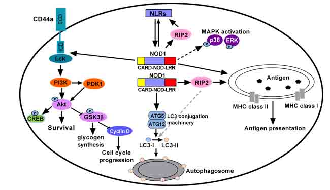
Receptor-Interacting Protein 2 (RIP2) is a key adaptor protein in NOD-like receptors (NLRs)-mediated signaling pathway. The in vitro function of RIP2 has been reported in teleost fish. However, the role and mechanisms of piscine Nucleotide Binding Oligomerization Domain (NOD)-RIP2 axis remain largely unknown in vivo.
Recently, the research group led by Prof. CHANG Mingxian from Institute of Hydrobiology (IHB) of Chinese Academy of Sciences obtained a knockout homozygous line of zebrafish NOD1 and RIP2. They investigated the effect of NOD1-RIP2 axis on immune signaling pathways during zebrafish early development by transcriptome sequencing and Western blotting.
Similar to NOD1, RIP2 deficiency significantly affected zebrafish hatching and larval survival. The significantly enriched pathways regulated by RIP2 were mainly involved in immune system, such as “Antigen processing and presentation”, “NOD-like receptor signaling pathway”, "Toll-like receptor signaling pathway” and “TNF signaling pathway” and so on.
Both NOD1 and RIP2 deficiency significantly impaired the expression of CD44a, however the downstream signaling of CD44a-Lck-PI3K-Akt pathway regulated by NOD1 was independent on RIP2. The regulation of NOD1 on the Mitogen-Activated Protein Kinase (MAPK) pathways was also independent on RIP2.
These findings highlight the similarity and discrepancy of NOD1 and RIP2 in the regulation of immune signaling pathways during the zebrafish early ontogenesis.
These results have been published in Frontiers in Immunology entitled “RIP2 is a critical regulator for NLRs signaling and MHC antigen presentation but not for MAPK and PI3K/Akt pathways”.